Suspecting that all was not well, a 42-year-old woman recently underwent a breast scan at one of Delhi’s leading hospitals. The scan revealed that she had a malignant tumour. Ordinarily, doctors would have conducted a surgery to conserve the breast, removing just the cancerous growth and not the entire breast. In this case, however, they opted for total mastectomy. That’s because the super-specialised scan that the woman was subjected to had not only detected the existing cancer, but it had also disclosed how the cancer would grow and extend in the future. It had caught the cancer’s field. And it had picked up three other cancer hotspots which a regular magnetic resonance imaging, or MRI, would have missed.
Called PET MRI, this specialised cancer diagnostic technology has changed the treatment line altogether by arming doctors with far more accurate data concerning cancer in their patients. It has also made it feasible to detect the possibility of a perfectly healthy individual having cancer in the future. “It can maybe pick up a cancer as early as five years or more,” says Shubham Sogani, the CEO of House of Diagnostics and PET Suite at Indraprastha Apollo Hospital in Delhi.
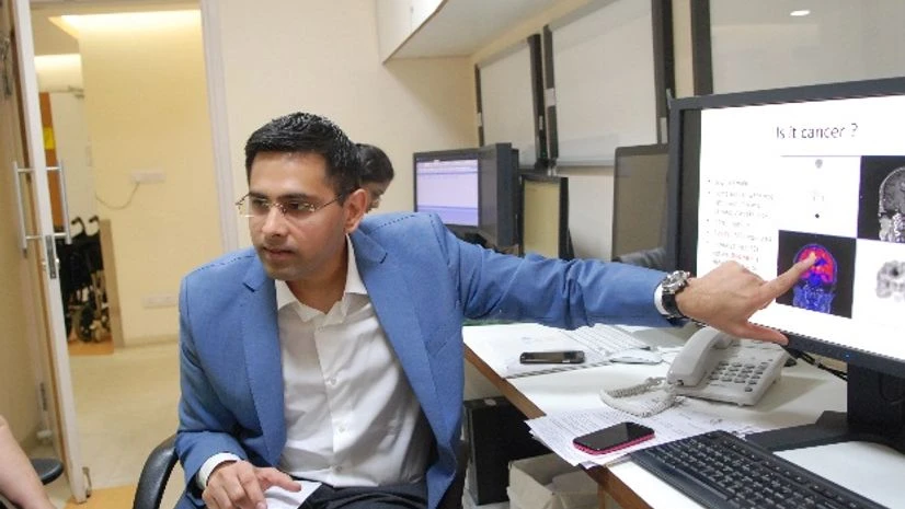
PET, or positron emission tomography, is a nuclear medicine imaging technique. Here is how it works. A biological radioactive material is injected into the body. The most commonly used radioactive material is FDG, or fluorodeoxyglucose, a glucose molecule. The patient is made to fast overnight so that “when he comes for the scan, he is starving and the body requires glucose,” says Sogani. The patient is now subjected to the radioactive glucose molecule. In about 45 minutes, the glucose is distributed throughout the body. Under the scan, anything that is normal shows a particular pattern of glucose uptake, while an anomalous pattern indicates an abnormality. That abnormality can be a cancer or an infection. “Normally, cancers and infections are rapidly multiplying cells. When anything multiplies rapidly, it needs more energy and, hence, its consumption of glucose is automatically higher,” explains Sogani. Based on this fundamental, the radioactive glucose molecules get concentrated in the problem area. When the patient is put under the scanner, the machine detects the high degree of radiation in that problem area. The intensity, direction and angle of the radiation hitting the scanner are picked up in 3D and the source is figured out. When this information is fused with an MRI scan, it gives the precise location of the cancer in the body. “PET tells us ‘yes, this is cancer’. And PET plus MRI tells us the specificity and extent of the cancer,” says Sogani.
| HOW OFTEN SHOULD YOU SCREEN FOR CANCER? |
|
|
There are over a thousand types of cancers. But routine whole-body checkups scan for only five or six types of prevalent cancers like breast, cervical, prostate or lung. In comparison, PET MRI, which is also a whole-body scan, picks up cancers or infections in any part of the body and with a greater precision.
The facility at Apollo is a simultaneous PET MRI. This means that the PET scan and MRI scan are conducted simultaneously and the data from the two is picked up simultaneously. Hence, the diagnosis is highly accurate. The person is also subjected to only one scan which lasts for 45 to 60 minutes.
| TINY WEAPONS, TINIER RISK |
|
THE SUPER MOLECULES NOT EVERY CANCER is picked up accurately by the FDG molecule. For a cancer or suspected cancer of the brain or spine, a unique super molecule called FET is used. After radiotherapy or surgery, the brain architecture alters. “If a tumour recurs now, it becomes very difficult to understand in which area and to what extent it has reappeared,” says Sogani. He cites the example of 49-year-old man who had undergone brain surgery and radiotherapy in 2009. Recurrence was suspected and the patient was subjected to both FET and FGD scans. While the glucose was detected in other normal areas of the brain as well, FET clearly showed where the recurrent tumour region lay. A precision surgery was conducted and tests revealed high-grade cancer. “The brain is such an area that you should do very limiting, specific surgery without disturbing the other areas,” says Sogani, “or else you can alter the patient altogether. You can paralyse him, vegetate him or even kill him.” After this, the patient was subjected to highly targeted radiotherapy so that normal tissue would not be affected. The new technology has equipped doctors to take a more scientific approach for a more effective line of treatment instead of shooting in the dark. It has also reduced patient trauma and cost of treatment. Another novel molecule used is DOTA. This is used for detecting neuroendocrine tumours, which can be found anywhere in the body, be it head, neck, thigh or spleen. These are all precious molecules. FET, for example, is produced at the Institute of Nuclear Medicine and Life Sciences and a seven-day notice is needed to procure it. FDG can be sourced from only two private vendors; one is in Noida and the other at Dera Bassi near Chandigarh. The half life of all these molecules is 110 minutes. This means that in 110 minutes, the radioactivity in these molecules get reduced by half. Hence, it is important that you reach the hospital at the designated time because the doctor might wait for you, but the molecule won’t. RISK OF RADIATION THE ATOMIC ENERGY Regulatory Board says that a person should be exposed to less than one millisievert of radiation in a year. So, how risky is PET MRI? Not too bad, it turns out. MRI emits no radiation because it is a magnetic test. So the total exposure in a PET MRI is solely because of the radioactive molecule that is injected. And that’s about 3 to 5 millisievert. “Compare that to a pilot who flies from London to the US in nine of the 12 months that he’s working. He is subjected to about 9 millisievert of cosmic radiation,” says Sogani. This makes PET MRI a safe test for picking up cancers in children and women of reproductive age. As opposed to PET MRI, PET CT scans involve greater exposure to radiation. The PET CT technology has been around in India for about two decades. Depending on the quality of the equipment, the radiation exposure from PET CT is about 15-30 millisievert. However, Choudhury of the Rajiv Gandhi Cancer Institute & Research Centre maintains that currently, PET CT is a better option. “You cannot replace PET CT with PET MRI, which is still an evolving technology worldwide. In PET CT, we know what to and what not to expect.” HOW MUCH DOES IT COST?
In India, the PET MRI test costs about one-seventh of what it does anywhere else in the world. In Singapore, for example, the test costs $S 4,000 (around Rs 2 lakh); in Europe, it costs euro 3,000-4,000 (Rs 2.3 lakh to Rs 3 lakh); and in the US, it is about Rs 4 lakh. This explains the number of patients from Europe, Africa and West Asia coming to Delhi for the test. |

)
